Public projects
OCT@home (planning)
Monitoring von Retinatherapien @HOME
The aim of the OCT@Home project is to improve the health care for wet AMD patients. To optimize injection therapy, an OCT for the home diagnosis is being developed at MLL. With OCT@HOME the patient can autonomously control disease progress and the necessary of a therapeutic intervention in in his own home.
Aim is to spared unnecessary injections and at the same time to avoid rapid reduction of vision by the increased control frequency.
Homecare of AMD
Age-related macular degeneration is a retinal disease that affects the area of sharpest vision. In Germany, about 1.6 million people are affected by AMD. It progresses batch-wise and, if left untreated, irreversibly damages the visual cells and thus leads to blindness. In Germany, AMD is responsible for every second blindness. The most important tool for diagnosing AMD is optical coherence tomography (OCT).
Age-related macular degeneration is a retinal disease that affects the area of sharpest vision. In Germany, about 1.6 million people are affected by AMD. It progresses batch-wise and, if left untreated, irreversibly damages the visual cells and thus leads to blindness. In Germany, AMD is responsible for every second blindness. The most important tool for diagnosing AMD is optical coherence tomography (OCT).
Meanwhile, wet AMD can be effectively treated. For this purpose, so-called VEGF inhibitors are injected by syringe into the eyeball of the patient. The best results are achieved with monthly injections. However, the procedure is relatively expensive at around 1500, – EUR per injection. In addition, the injections into the eyeball are very stressful for many patients.
As a result, less frequent injection schemes are now being used to reduce the number of injections. However, these have the disadvantage that due to the extended examination intervals, a new disease relapse is often recognized too late. The situation could be improved with more frequent OCT examinations (for example at weekly or even daily intervals). However, the effort required for this exceeds the capacity of the medical infrastructure and is not feasible for most patients.
The alternative to the existing scheme is self-examination by the patient with an OCT device that he can be used at home, much like a sphygmomanometer. This allows a daily examination interval. Thus, the doctor and patient would be warned at the beginning of a new episode of the disease and therefore have enough time to prevent a loss of vision by an additional injection.
Technology
Since mid-2015, the MLL is working on an OCT procedure suitable for home diagnosis as part of the BMBF-funded research project RETOME. For this purpose, a new, previously unknown measuring principle for the acquisition of OCT data was used, which was developed at the Institute of Biomedical Optics at the University of Lübeck.
The technology, which for systematic reasons is called Off-Axis-Full-Field Time-Domain OCT, differs in essential points from the FD-OCT technique used in everyday clinical practice:
- Instead of using a high-resolution spectrometer, the different depths in the sample are scanned by the stepwise displacement of a small mirror.
- Instead of scanning the retina point by point with galvanometers, a complete plane is captured with a single acquisition using a commercially available image sensor.
- Instead of compensating the ametroptia of the patient by shifting optomechanical components, this is compensated numerically.
- Complex fibre optic components are replaced by simple and much cheaper bulk optics.
A detailed description of the process and the scientific background can be found here.
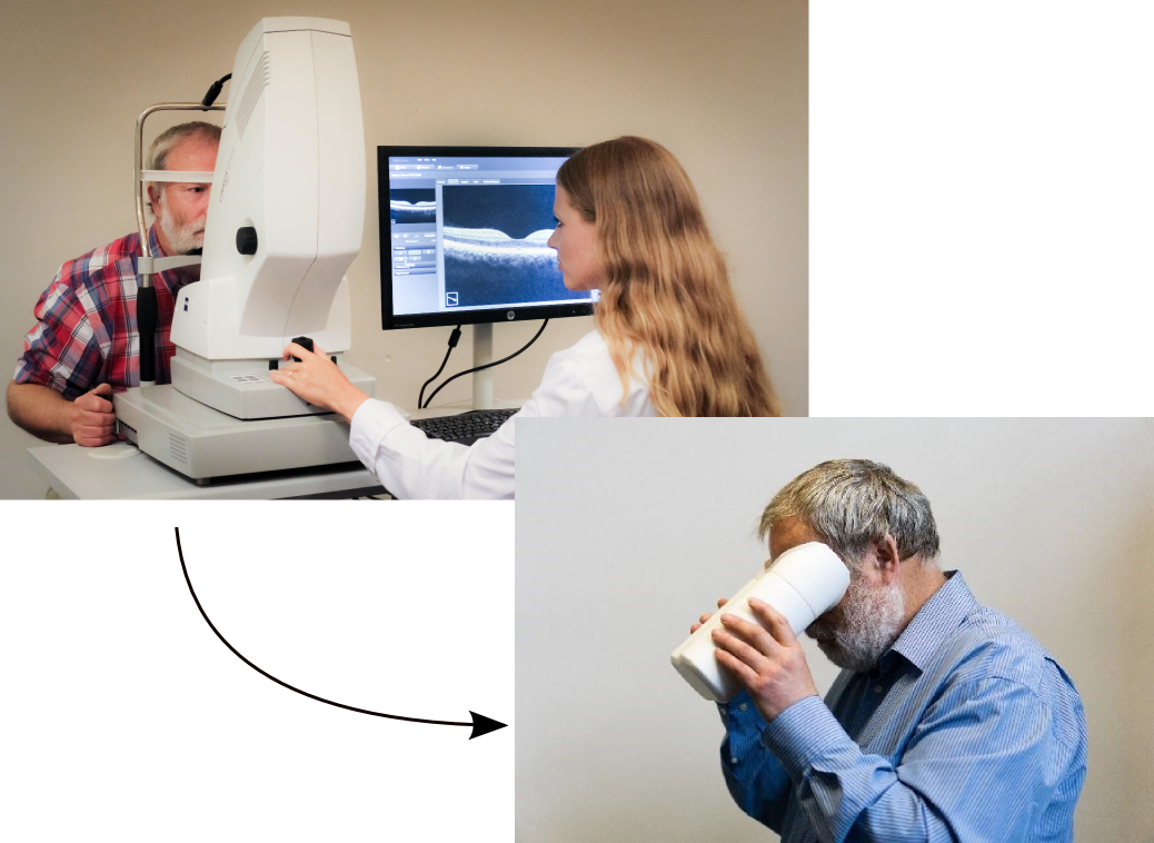
In contrast to previous OCT devices, all optical components can be integrated in a handheld device. A fully functional demonstrator was realised and tested on volunteers.
Clinical Evaluation
Clinical Evaluation of the home-care OCT is currently conducted in the context of the BMBF project RETOME at the Eye Clinic of the University of Kiel. By June 2018, 10 patients with different pathologies were included.
- Even patients with a residual visual acuity of only 5% were able to operate the device on their own.
- The image quality does not yet reach that of the FD-OCT reference measurements, but is still suitable for AMD diagnostics.
- Image quality was only marginally degraded by typical previous illnesses (eg slight cataract).
Research
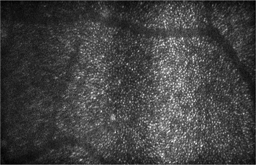
The technique shown here can also be used to numerically correct aberrations in the imaging optics and the eye itself. This allows the visualization of individual photoreceptor cells of the retina. This has previously been possible only with significant more complex OCT systems based on adaptive optics.
This research is done in close collaboration with the Institute of Biomedical Optics of the University of Lübeck (PD Dr. Gereon Hüttmann).