public Projects
Project Holo-OCT
Computer assisted Holographic OCT
Eye diseases that can lead to loss of vision are becoming increasingly common – with dramatic consequences for those affected. Optical technologies for imaging the retina have contributed decisively to advances in the diagnosis and treatment of retinal diseases. In particular, optical coherence tomography (OCT), which, like ultrasound, uses the time-of-flight differences of waves for depth resolution, has become indispensable for the successful treatment of common diseases such as age-related macular degeneration by visualising the structure of the retina. New therapies in ophthalmology require methods that also provide functional information at the cellular level.
Optical coherence tomography (OCT) is one of the standard diagnostic methods in ophthalmology. In addition to the spatially resolved measurement of the intensity of the backscattered light, OCT also provides access to the phase of the light. In principle, this enables holographic imaging, which can numerically correct image errors and measure morphological changes in tissue with nanometre precision. However, the phase information is strongly disturbed by eye movements and is not yet usable in the clinic. In particular, holographic OCT enables functional measurements on individual cells of the retina. As optical retinography (ORG), it has the potential to map visual and neuronal function of the retina non-invasively with high spatial accuracy and thus contribute to early detection of important ophthalmological and neuronal diseases. In the project Holo-OCT, the phase of the OCT signal is to be made usable for clinical diagnostics by increasing the imaging speed and numerically correcting the movements.
An evaluation of holographic OCT is only possible to a limited extent in phantoms and animal models, since neither the physiological processes of the retina nor the imaging conditions with patients (e.g. movements and pathological anatomy of the eye) can be realistically represented. For a validation of the holographic OCT, measurements on test persons and patients within a clinical trial are necessary.
The MLL realises a functional demonstrator, which can be clinically applied according to Art. 82 of the Medical Device Regulation (MDR). For this purpose, Holo-OCT images are first taken on healthy test persons. After scientific analysis of these images, the Holo-OCT system will be evaluated in a clinical study.
The scientific evaluation of the study is carried out in cooperation with the project partners of the Charité -Universitätsmedizin Berlin (ECRC) and the Institute for Biomedical Optics at the University of Lübeck (BMO).

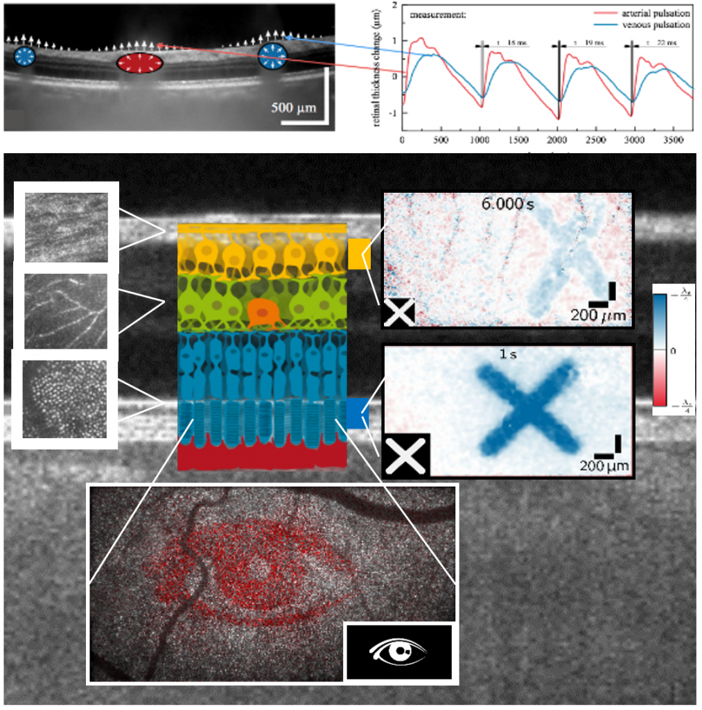
Cellular-resolved functional retinal imaging with holographic OCT.
a) Interferometric measurement of the distance between two layers of the retina allows tracking of pulse waves in arteries and veins.
b) High-resolution imaging of nerve fibres, capillaries and individual photoreceptors by digital adaptive optics (DAO) (left).
Evaluation of phase differences across the outer segments (blue) or ganglion cells (yellow) makes reaction of these cells after optical stimulation visible (right).
Combination with DAO enables functional measurements on individual cells (bottom).