Public projects
UltraLas
Optical measurement techniques combined with ultrasound or laser tissue resection in neurosurgery for local detection of tissue boundaries, elasticity and vascular architecture
Optimized tumor resection in neurosurgery
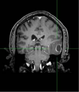
In Germany, about 43,000 new oncological diseases are diagnosed each year in the central nervous system (CNS). This number will continue to increase in the future due to demographic trends. Microsurgical resection is the standard treatment for most of the tumors in the CNS. The survival rate depends, among other things, on the extent of resection. To date, however, detection of tumor margins versus intact tissue intraoperatively is difficult, or it can only be accomplished with great technological effort. Another problem is the strongly fluctuating dissection rate in the use of ultrasound or laser instruments and the lack of visibility of the underlying vessel courses. The result is a difficult choice for the surgeon in terms of device parameters and intraoperative bleeding, which are currently mostly stopped with current-carrying bipolar forceps. Disadvantages of this contact-based hemostasis include among other problems due to tissue adhesions at the electrodes and an induced even more difficult discrimination of tumorous tissue by color / haptic. The overall goal of the collaborative work is therefore to improve the resection extent during the operation by the automatic detection of tumor properties and tumor margins. At the same time, the operation associated risk to the patient should be minimized, leading to an extended lifetime with a high quality of life.
Optical measurement methods combined with ultrasound or laser tissue resection for clinical practice
In this project, various methods for intraoperative in vivo detection of tumor expansion, vascular architecture and tumor elasticity in neurosurgery are to be developed and evaluated. Several innovative photonic methods one the hand are to be used for a rapid and non-invasive measurement of the lesions, and on the other hand, should lead to effective dissection and coagulation of the tumor tissue. Sub-goals are novel system solutions of optical measuring methods in combination with laser or ultrasonic instruments. Such a combination of different technologies combines intraoperative, comparatively inexpensive optomechanical tissue analysis with high local resolution and visualization of deep vascular architecture with an improved laser or ultrasound instrument. This novel optimization of the resection procedure enables a significantly improved tumor resection in clinical practice through supportive resection margin recognition and optimized dissection performance. After completion of the project, the results should be converted into marketable products.
Paper:
Proceedings
Sazgar Burhan, et al., Phase analysis strategies for MHz OCE in the large displacement regime,
Sazgar Burhan et al., Advanced FFT-based contrast approach for MHz optical coherence elastography,
Hutfilz, A. Laser application for non-destructive blood vessel haemostasis on brain
tissue as an alternative to bipolar forceps, DGNCH 2022
Hutfilz A, et al. Laser coagulation of brain tissue at 1480 nm and 1940 nm wavelengths: SPIE; 2021.
J.Kren et al., Hand-guided robotic assistance for stabilized intraoperative diagnostic probe handling in brain tumor surgeries, RC301, DGNC Congress 2026, Aachen, Germany
J.Kren et al., Mechanical Tissue Properties as Markers for Brain Tumor Tissue Characterization, RC309, DGNC Congress 2026, Aachen, Germany